
Sonography, High-Resolution 3D/4D Obstetric Scans & Pelvic Doppler
Immediate, high-precision diagnostic ultrasound personally conducted and interpreted by Dr. M. Ananda Kumari, delivering instant clinical clarity for expecting mothers and gynaecology patients in Vijayawada.
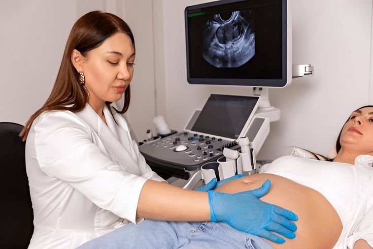
High-Resolution Pelvic Ultrasound, Obstetric Sonography & Color Doppler
Diagnostic Sonography is the gold-standard, radiation-free imaging modality in modern obstetrics and gynecology. Utilizing high-frequency acoustic wave probes, Color Flow Doppler, and 3D volumetric surface rendering, ultrasound allows real-time visualization of pelvic organ morphology, endometrial architecture, and fetal morphogenesis. At DR. AK CLINIC, clinical ultrasound is conducted and directly interpreted by senior gynecologist Dr. M. Ananda Kumari, ensuring immediate clinical correlation without the disconnect or delays of external scanning centers.
Waiting for an ultrasound scan or trying to decipher technical diagnostic reports from an external lab causes immense anxiety for pregnant mothers and gynecological patients. By performing high-resolution sonography in-clinic, Dr. Ananda Kumari discusses the live ultrasound images with you on the monitor screen in real-time, explaining findings instantly and initiating immediate treatment if required.
Our range of services include :
Our patient-focused approach integrates precision diagnostics, advanced 3D keyhole surgery, and 25+ years of clinical excellence for superior recovery and long-term well-being
Over 25 years of specialized surgical excellence in Vijayawada led by Dr. M. Ananda Kumari
Trusted by generations of women across Andhra Pradesh for compassionate, personalized gynaecological care
Extensive surgical track record in 3D laparoscopic, hysteroscopic & preventive oncology procedures
Dedicated to medical excellence, rapid recovery, and patient-centered clinical care



Transvaginal sonography confirming gestational sac implantation, fetal heart motion, precise gestational age, and ruling out ectopic pregnancy.
High-resolution 11–13.6 weeks ultrasound measuring Nuchal Translucency and nasal bone to assess early fetal chromosomal health.
Detailed blood-flow resistance indexing of uterine, umbilical, and ovarian vessels to diagnose preeclampsia risk, cysts, and tumors.
Serial ultrasound measuring follicle diameter and multi-layer endometrial thickness to identify the optimal fertility window.
Detailed Medical Conditions, Warning Signs & Evidence-Based Care
First-trimester spotting or bleeding, sudden unilateral pelvic pain (concerning for ectopic pregnancy or ovarian cyst rupture/torsion), missed periods, abnormal uterine bleeding, pelvic pressure, or reduced fetal movement in pregnancy.
Accurate dating of pregnancy, detecting multiple gestations (twins), screening for placental anomalies (placenta previa/accreta), mapping uterine fibroid numbers and depths before surgery, and monitoring follicular rupture for IUI.
Transabdominal Ultrasound (TAS) for wide pelvic overview, high-frequency Transvaginal Ultrasound (TVS) providing crisp millimeter-level resolution, 3D surface reconstruction, and real-time Color Flow Doppler mapping.

Direct consultation with Dr. M. Ananda Kumari immediately after the scan, eliminating anxious waiting times, external lab travel, or miscommunicated reports, followed by immediate medical or surgical planning.
Urgent Symptoms Requiring Immediate Sonographic Evaluation
Certain obstetric and gynaecological symptoms are time-critical medical situations where prompt ultrasound diagnosis protects maternal and fetal safety.
- First-Trimester Bleeding with Pelvic Cramping – Any spotting or bleeding in early pregnancy requires immediate ultrasound confirmation of fetal viability and ruling out threatened miscarriage.
- Severe One-Sided Pelvic Pain with Positive Pregnancy Test – Sharp, intense unilateral pain that may cause fainting or dizziness — an immediate emergency requiring ruling out an ectopic pregnancy.
- Sudden Decrease or Cessation of Fetal Movements – Noticeable reduction in daily fetal kick counts during late second or third trimester demands immediate biophysical profile and Doppler flow check.
- Acute Sudden Lower Abdominal Pain in Non-Pregnant Women – Sudden, severe pelvic pain indicating possible ovarian cyst torsion (twisting cutting off blood supply) or internal cyst hemorrhage.
- Sudden Watery Fluid Leakage During Pregnancy (PPROM) – Premature rupture of membranes requiring immediate measurement of amniotic fluid levels (AFI) and fetal heart rate monitoring.
These symptoms require thorough clinical evaluation, precision imaging, and early intervention to rule out complications. Dr. M. Ananda Kumari provides advanced diagnostic consultations and prompt surgical/medical management at our Vijayawada centre.
Frequently Asked Clinical Questions
Scientifically verified medical guidance from Dr. M. Ananda Kumari regarding Sonography (Ultrasound).
At commercial scanning centers, scans are often performed by technicians, and patients must wait days for a typed report, leading to immense anxiety. At DR. AK CLINIC, Dr. M. Ananda Kumari personally conducts and interprets the scan in real-time, explaining findings to you immediately and prescribing treatment or reassurance on the spot with zero delay.
The first viability and dating scan is typically performed between 6 to 8 weeks of pregnancy. It confirms whether the pregnancy is safely located inside the uterus (ruling out dangerous ectopic pregnancy), detects the fetal heartbeat, confirms single or twin gestation, and calculates your precise expected due date (EDD).
The NT scan is a critical genetic ultrasound performed strictly between 11 weeks and 13 weeks 6 days of pregnancy. It measures the clear fluid space at the back of the baby's neck (nuchal translucency) and evaluates the fetal nasal bone to assess the risk of chromosomal conditions such as Down syndrome and early cardiac anomalies.
Yes, completely safe. Diagnostic medical ultrasound does not use ionizing radiation (unlike X-rays or CT scans); it uses high-frequency sound waves. Decades of clinical evidence and international medical bodies (ACOG, ISUOG) have confirmed that obstetric ultrasound performed by experienced physicians has no harmful effects on mother or baby.
Follicular monitoring is a series of 3 to 5 quick transvaginal ultrasound scans during a menstrual cycle (usually starting around Day 9 or 10) to track the growth of ovarian follicles (measuring 2mm per day) and endometrial thickness. It determines the exact day of peak ovulation to time natural intercourse or Intrauterine Insemination (IUI) for maximum conception success.